- Visibility 52 Views
- Downloads 5 Downloads
- DOI 10.18231/j.ijcbr.2022.058
-
CrossMark
- Citation
Evaluation of serum protein electrophoresis in patients with renal disorders
- Author Details:
-
Shiv Sharma *
-
Divya Soin
-
Harvinder Singh
-
Khushdeep Singh
Introduction
Electrophoresis is a method of separating proteins based on their physical properties. The serum is placed on a specific medium, and a charge is applied. The net charge (positive or negative) and the size and shape of the protein commonly are used in differentiating various serum proteins. There are several ways used to separate and differentiate various serum components. Major methods are zone electrophoresis and capillary electrophoresis.[1]
Components of serum proteins
The measurement of protein is done on serum, which is the fluid that remains after plasma has clotted, thus removing fibrinogen and most of the clotting factors. By measuring the concentration of these proteins, the clinician can obtain information regarding disease states in different organ systems. The normal serum protein level is 6 to 8 g/dl. Albumin makes up 3.5 to 5.0 g/dl, and the remainder is the total globulins. These values may vary according to the individual laboratory.[2]
Albumin
The albumin band represents the largest protein component of human serum. The albumin level is decreased under circumstances in which there is less production of the protein by the liver or in which there is increased loss or degradation of this protein. Malnutrition, significant liver disease, renal loss [e.g., in nephrotic syndrome], hormone therapy, and pregnancy may account for a low albumin level. Burns also may result in a low albumin level.
Alpha fraction
Moving toward the negative portion of the gel [i.e., the negative electrode], the next peaks involve the alpha1 and alpha2 components. The alpha1-protein fraction is comprised of alpha1-antitrypsin, thyroid-binding globulin, and transcortin. Malignancy and acute inflammation [resulting from acute-phase reactants] can increase the alpha1-protein band. A decreased alpha1-protein band may occur because of alpha1- antitrypsin deficiency or decreased production of the globulin as a result of liver disease. Ceruloplasmin, alpha2-macroglobulin, and haptoglobin contribute to the alpha2-protein band. The alpha2 component is increased as an acute-phase reactant.
Beta fraction
The beta fraction has two peaks labeled beta1 and beta2. Beta1 is composed mostly of transferrin, and beta2 contains beta-lipoprotein. IgA, IgM, and sometimes IgG, along with complement proteins, also can be identified in the betafraction.
Gamma fraction
Much of the clinical interest is focused on the gamma region of the serum protein spectrum because immunoglobulins migrate to this region. It should be noted that immunoglobulins often can be found throughout the electrophoretic spectrum. C- reactive protein [CRP] is located in the area between the beta and gamma components.[3]
Separated proteins
Human serum proteins have 70,000 to 250,000 molecular weight range. After staining, the bands are well-defined, the molecular weight is defined and, hence, the identity of each can be estimated from the migration distance.
Clinical significance of serum protein electrophoresis
The kidney is the major site of catabolism for small serum proteins. The relationship of normal renal function to the metabolism of serum proteins is reflected in the fact that proteinuria and abnormalities of serum protein concentration accompany most forms of renal disease.[4] The relationship of relative renal clearances of individual protein fractions to their corresponding molecular weight, serving as an index of glomerular permeability, is applied in the clinical investigation of patients with the nephrotic syndrome.[5] The functional defect in nephrotic syndrome is the severe urinary loss of protein and characterization of serum and urine proteins has been recognized to be of fundamental importance in the investigation of this condition.[6]
Plasma protein levels display reasonably predictable changes in response to acute inflammation, malignancy, trauma, necrosis, infarction, burns, and chemical injury. Monoclonal gammopathies are associated with a clonal process that is malignant or potentially malignant, including multiple myeloma, Waldenström’s macroglobulinemia, solitary plasmacytoma, smoldering multiple myeloma, monoclonal gammopathy of undetermined significance, plasma cell leukemia, heavy chain disease, and amyloidosis.[7]
The author planned the study to evaluate serum proteins using SDS PAGE method in Nephrotic syndrome patients. Chronic kidney patients and end stage renal disorders.
Aims and Objectives
The aim of the present study was to evaluate protein electrophoresis pattern in various renal disorders. i.e Nephrotic syndrome, end stage renal disorder and chronic kidney disease and were statistically analysed.
Materials and Methods
The present hospital-based observational study was conducted over a period of one year in department of Biochemistry on a total of 50 patients with renal disorder out of which 18 patients were with nephrotic syndrome, 21 were with ESRD, 11 were with CKD of either sex visiting the OPD/IPD of Medicine Department of Guru Gobind Singh Medical College and Hospital Faridkot were enrolled in the study and the patients of pregnancy, chronic infections, Cancer, smokers, malnutrition were excluded from the study.
Sampling technique/method
A sample of the patients with the renal disease were taken for study. 3 ml of the venous blood sample was drawn from each subject under aseptic condition. The blood sample was taken in plain vacutainer for biochemical investigation. After the formation of blood clot, the sample was centrifuged at 3000 rpm for 10 minutes to separate serum. After that we analyzed serum for the biochemical investigation.
The investigations required i.e Serum Creatinine, Blood Urea and Uric Acid had been done on the fully autoanalyser AU480 and for Serum protein electrophoresis SCIEPLAS vertical electrophoresis was used.
Prepration of Gels
|
Running gel (10%) |
|
|
Requirements for Running gel |
Quantity |
|
30% acrylamide |
3.4 ml |
|
dH2O |
4.0 ml |
|
1.5M TRIS, pH= 8.8 |
2.5 ml |
|
0.4% SDS |
100µl |
|
TEMED |
15 µl |
|
10% Ammoniium Perrssullffatte |
50 µl |
|
Stacking gel (4%) |
|
|
Requirements for Stacking gel |
Quantity |
|
30% acrylamide |
670 µl |
|
dH2O |
3.6 ml |
|
0.5M TRIS, pH= 6.8 |
630 µl |
|
10% SDS |
50 µl |
|
TEMED |
10 µl |
|
10% Ammonium Persulfate |
25 µl |
|
Preparation of sample buffer (50ml) |
|
|
Component |
2X |
|
TRIS pH=6.8 |
0.125 gm |
|
SDS |
2 gm |
|
2-ME² |
5 ml |
|
Glycerol |
10 ml |
|
Bromophenol brilliant blue (0.1%) |
0.1 gm |
Sample preparation
3.5 µl of serum sample was added to 3.5 µl sample buffer. Sample was then heated up for denaturation and then added to sample wells.
In stacking gel voltage was 60 V.
In resolving gel voltage was 120 V
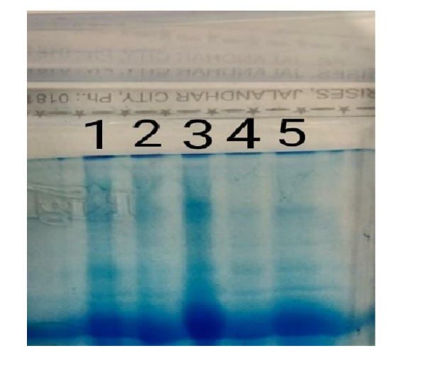
After run, staining was done by commassie brilliant blue G-250.
Staining with Coomassie Blue R250
Gel was stained with 0.1% (or less) Coomassie Blue R250 in 10% acetic acid, 50% methanol, and 40% H2O for the minimum time (typically one hour) necessary to visualize the bands of interest.
Then it was destained by soaking for at least 2 hours in 10% acetic acid, 50% methanol, and 40% H2O with at least two changes of this solvent. If the gel had Coomassie Blue background then destaining was continued until the background is nearly clear.
After that the stained gel was placed in GelDoc (SyngeneGbox) and image was captured. The image was further read by particular software.
The amount of protein was calculated by following formula:-
Amount of protein content = 𝐵𝐴𝑁𝐷% × 𝑇. 𝑃.
(Where, a) BAND% = Percentage of Band in particular lane
b) T.P = Total Protein)
Statistical analysis
The data was analysed using Microsoft excel 7 and was done on SPSS20.
Results
|
Band % |
NS |
ESRD |
CKD |
|
Albumin |
41.26±5.7 |
48.3±4.9 |
51.3±3.5 |
|
Apha 1 |
4.6±0.62 |
5.6±1.31 |
4.41±0.87 |
|
Alpha 2 |
15.2±3.3 |
11±2.3 |
11.8±2.4 |
|
Beta |
17.5±3.54 |
13.9±3.21 |
14.6±2.57 |
|
Gamma |
20±4.71 |
21.9±6.9 |
22.3±4.1 |
[Table 4] shows Mean and standard deviation of Band% in patients having nephrotic syndrome, end stage renal disorder, chronic kidney disease. Band % is percentage of particular band in whole run electrophoretic pattern (Data represented as mean±SD).
|
|
Mean±SD of NS |
Range |
Mean±SD of ESRD |
Range |
Mean±SD of CKD |
Range |
|
Albumin (gm/dl) |
2.20±0.62 |
1.58-2.82 |
2.90±0.51 |
2.39-3.41 |
3.34±0.40 |
2.94-3.74 |
|
Apha 1 (gm/dl) |
0.23±0.03 |
0.20-0.26 |
0.34±0.07 |
0.27-0.41 |
0.28±0.05 |
0.23-0.33 |
|
Alpha 2 (gm/dl) |
0.78±0.16 |
0.62-0.94 |
0.66±0.11 |
0.55-0.77 |
0.71±0.13 |
0.58-0.84 |
|
Beta (gm/dl) |
0.9±0.15 |
0.75-0.24 |
0.82±0.18 |
0.64-1 |
0.88±0.13 |
0.75-1.01 |
|
Gamma (gm/dl) |
1.10±0.26 |
0.84-1.36 |
1.28±0.49 |
0.79-1.77 |
1.31±0.34 |
0.97-1.65 |
[Table 5] shows Fraction of proteins in patients having nephrotic syndrome, end stage renal disorder, chronic kidney disease by serum protein electrophoresis (Data represented as mean±SD).
|
|
NS |
ESRD |
t value |
p-value |
Significance |
|
Albumin (gm/dl) |
2.20±0.62 |
2.90±0.51 |
3.86 |
<0.05 |
Significant |
|
Apha 1 (gm/dl) |
0.23±0.03 |
0.34±0.07 |
6.18 |
<0.05 |
Significant |
|
Alpha 2 (gm/dl) |
0.78±0.16 |
0.66±0.11 |
-2.76 |
<0.05 |
Significant |
|
Beta (gm/dl) |
0.9±0.15 |
0.82±0.18 |
-1.49 |
0.14 |
Not significant |
|
Gamma (gm/dl) |
1.10±0.26 |
1.28±0.49 |
1.39 |
0.17 |
Not significant |
[Table 6] of our study shows Mean±SD of Albumin, α1 Globulin, α2 Globulin, ß Globulin and Gamma Globulin in case of NS and ESRD. The data shows the high significant values (p<0.05) in case of Albumin (t=3.86),α1 Globulin(t=6.18), α2 Globulin (t=-2.76) whereas, for ß Globulin and Gamma Globulin values calculated were not significant (Data represented as mean±SD).
|
|
NS |
CKD |
t value |
p-value |
Significance |
|
ALBUMIN (gm/dl) |
2.20±0.62 |
3.34±0.40 |
5.42 |
<0.05 |
Significant |
|
APHA1 (gm/dl) |
0.23±0.03 |
0.28±0.05 |
3.38 |
<0.05 |
Significant |
|
ALPHA2 (gm/dl) |
0.78±0.16 |
0.71±0.13 |
-1.22 |
0.23 |
Not significant |
|
BETA (gm/dl) |
0.9±0.15 |
0.88±0.13 |
-0.36 |
0.71 |
Not significant |
|
GAMMA (gm/dl) |
1.10±0.26 |
1.31±0.34 |
1.87 |
0.07 |
Not significant |
[Table 7] of our study shows the mean and standard deviation of Albumin, α1 Globulin, α2 Globulin, ß Globulin and GammaGlobulin in case of Nephrotic Syndrome and Chronic Kidney Disease. The data shows the high significant values (p<0.05) in case of Albumin(t=5.42), α1 Globulin (t=3.38) whereas, for α2 Globulin, ß Globulin and Gamma Globulin values calculated were not significant (Data represented as mean±SD).


Discussion
Gel electrophoresis is widely known technique used to separate and identify macromolecules. There are many types of gel electrophoresis, sds page technique is used to characterize complex complex protein mixtures. The proteins have net electrical charge if they are in medium having pH different from isoelectric point and have ability to move when subjected to an electric field. The different type of serum proteins are:
Albumin. Albumin proteins keep the blood from leaking out of blood vessels. More than half of the protein in blood serum is albumin.
Alpha-1 globulin. High-density lipoprotein [HDL], the "good" type of cholesterol, is included in this fraction.
Alpha-2 globulin. A protein called haptoglobin, which binds with hemoglobin, is included in the alpha-2 globulin fraction
Beta globulin. Beta globulin proteins help carry substances, such as iron, through the bloodstream and help fight infection.
Gamma globulin. These proteins are also called antibodies. They help prevent and fight infection.
Each of these five protein groups moves at a different rate in an electrical field and together form a specific pattern. This pattern helps identify some diseases.
The SDS-PAGE in combination with a protein stain is widely used in biochemistry for the quick and exact separation and subsequent analysis of proteins. In medical diagnostics, SDS-PAGE is used as part of the HIV testand to evaluate proteinuria. In the HIV test, HIV proteins are separated by SDS-PAGE and subsequently detected by Western Blot with HIV-specific antibodiesof thepatient.
Therefore, the aim of the present study was to evaluate the role of serum protein electrophoresis in patients with renal disorder.
The present study was conducted in the Department of Biochemistry. A total of 50 individuals with renal patients were enrolled in the study in which 33 were males and 17 were females.
[Table 4] of our study shows the mean and standard deviation of Band % of Albumin(gm/dl), α1 Globulin(gm/dl), α2 Globulin(gm/dl), ß Globulin(gm/dl) and Gamma Globulin(gm/dl). Values of Band% in case of ESRD were found to be 48.3±4.9, 5.6±1.31, 11±2.3, 13.9±3.21, 21.9±6.9. In case of nephrotic syndrome Values of Band% were found to be 41.26±5.7, 4.6±0.62, 15.2±3.3, 17.5±3.54 and 20±4.71 respectively. The CKD patients Value of Band % were found tobe 51.3±3.5, 4.41±0.87, 11.8±2.4, 14.6±2.57 and 22.3±4.1.
[Table 5] of our study shows the mean and standard deviation of protein fraction of Albumin(gm/dl), α1 Globulin(gm/dl), α2 Globulin(gm/dl), ß Globulin(gm/dl) and Gamma Globulin(gm/dl). In case of Nephrotic Syndrome was found to be 2.20±0.62, 0.23±0.03, 0.78±0.16, 0.9±0.15 and 1.10±0.26 respectively. Similarly, values in case of ESRD values were 2.90±0.51, 0.34±0.07, 0.66±0.11, 0.82±0.18 and1.28±0.49. In CKD values were 3.34±0.40, 0.28±0.05, 0.71±0.13, 0.88±0.13 and 1.31±0.34.
[Table 6] of our study shows the comparison of protein content in patients of NS and ESRD at p<0.05. Mean and standard deviation of Albumin(gm/dl), α1 Globulin(gm/dl), α2 Globulin(gm/dl), ß Globulin (gm/dl) and Gamma Globulin(gm/dl) in case of Nephrotic Syndrome were found to be 2.20±0.62, 0.23±0.03, 0.78±0.16, 0.9±0.15 and 1.10±0.26 respectively. Similarly, values in case of ESRD values were 2.90±0.51, 0.34±0.07, 0.66±0.11, 0.82±0.18 and1.28±0.49. The data shows the high significant values (p<0.05) in case of Albumin (t=3.86), α1 Globulin(t=6.18), α2 Globulin (t=-2.76) whereas, for ß Globulin and Gamma Globulin values calculated were not significant(p>0.05).
table of our study shows the comparison of protein content in patients of NS and CKD at p<0.05. Mean and standard deviation of Albumin (gm/dl), α1 Globulin(gm/dl), α2 Globulin(gm/dl), ß Globulin (gm/dl) and Gamma Globulin(gm/dl) in case of Nephrotic Syndrome was found to be 2.20±0.62, 0.23±0.03, 0.78±0.16, 0.9±0.15 and 1.10±0.26. Similarly, values in case of CKD values were 3.34±0.40, 0.28±0.05, 0.71±0.13, 0.88±0.13 and 1.31±0.34. The data shows the high significant values (p<0.05) in case of Albumin (t=5.42), α1 Globulin (t=3.38) whereas, for α2 Globulin, ß Globulin and Gamma Globulin values calculated were not significant (p>0.05).
In case of NS, because of the size of α-2 macroglobulin is retained in the bloodstream while albumin levels are decreased.
In case of ESRD, hypoalbuminemia occurs and due to inflammatory response levels of α-1 globulin increased.[8]
Conclusion
The study concluded that serum protein fractions are altered in various renal disorders. Protein electrophoresis should be used in addition to other renal markers foe better monitoring of the disease progression.
Source of Funding
None.
Conflict of Interest
None.
References
- RF Jacoby, CE Cole. Molecular diagnostic methods in cancer genetics. Int J Biol Med Res 2015. [Google Scholar]
- JT Busher, HK Walker, WD Hall, JW Hurst. . Clinical Methods the History, Physical, and Laboratory Examinations 1990. [Google Scholar]
- M Al Hiary, A Momani, R Khasawneh, B Zghoul, L Habahbeh, AR Wreikat. Patterns of serum protein electrophoresis. Int J Biol Med Res 2015. [Google Scholar]
- TA Waldmann, W Strober, RP Mogielnicki. The Renal Handling of Low Molecular Weight Proteins. J Clin Investig 1972. [Google Scholar]
- JD Blainey, BD Brewer, JH Hardwicke, JF Soothill. The nephrotic syndrome. Diagnosis by renal biopsy and biochemical and immunological analyses related to the response to steroid therapy. Quart J Med 1960. [Google Scholar]
- DS Rowe. The molecular weights of the proteins of normal and nephrotic sera and nephrotic urine, and a comparison of selective ultrafiltrates of serum proteins with urine proteins. Biochem J 1957. [Google Scholar]
- WC Griffiths, HC Boqaars, CF Sheehan, BS Diamond. Analysis of human serum proteins by molecular weight dependent acrylamide gel electrophoresis. Ann Clin Lab Sci 1976. [Google Scholar]
- N Takei, M Suzuki, H Makita, S Konno. Serum alpha-1 Antitypsin levels and the clinical course of chronic obstructive pulmonary disease. Int J Chron Obstruct Pulmon Dis 2019. [Google Scholar]
- Introduction
- Components of serum proteins
- Albumin
- Alpha fraction
- Beta fraction
- Gamma fraction
- Separated proteins
- Clinical significance of serum protein electrophoresis
- Aims and Objectives
- Materials and Methods
- Sampling technique/method
- Prepration of Gels
- Sample preparation
- Staining with Coomassie Blue R250
- Statistical analysis
- Results
- Discussion
- Conclusion
- Source of Funding
- Conflict of Interest
